Products
Categories
Educational Equipments

LUMBAR VERTEBRAE, PELVIS AND SACRUM WITH NERVES
LUMBAR VERTEBRAE, PELVIS AND SACRUM WITH NERVES

MAGNIFIED ATLAS MODEL WITH PLASTIC BASE
MAGNIFIED ATLAS MODEL WITH PLASTIC BASE

Magnified Human Larynx Model
A functional model that demonstrates movements of the epiglottis and cartilages in the voice box. It helps the students to require and understanding of the morphology and structure of the respiratory tract and phonetic organ. On base. 3 times enlarged. 3- parts dissectible. Available in Packing: 62 x 29 x 29 cm, 12 pcs/carton, 10 kgs.

MAGNIFIED HUMAN VENTRICLES AND BASAL NUCLEI MODEL

Magnified Pulmonary Alveoli Model
The model shows the small branches of principal bronchus:
Section of bronchiole of no cartilage. The relation between pulmonary alveoli and terminal bronchiole. The structure of alveolar sac and alveolar duct.
The capillary rete in the alveolar sapta. Made of PVC plastic and mounted on plastic base in Size: 26x15x35 cm.
Section of bronchiole of no cartilage. The relation between pulmonary alveoli and terminal bronchiole. The structure of alveolar sac and alveolar duct.
The capillary rete in the alveolar sapta. Made of PVC plastic and mounted on plastic base in Size: 26x15x35 cm.

MAGNIFIED THALAMIC MODEL WITH BASE

MAGNIFIED TRANSET OF SPINAL MARROW SHOWING THE: RELATION
MAGNIFIED TRANSET OF SPINAL MARROW SHOWING THE: RELATION

Magnified Uterus Model
With the coronal section this model shows:
1. Uterus
2. The cervix is spanided into a supra-vaginal portion
and a vaginal portion.
3. The ligaments of the uterus are eight in number.
4. The fallopian tubes
5. The ovaries
Available in Packing: 50 x 35 x 40 cm, 20 pcs/carton, 15 kgs.
1. Uterus
2. The cervix is spanided into a supra-vaginal portion
and a vaginal portion.
3. The ligaments of the uterus are eight in number.
4. The fallopian tubes
5. The ovaries
Available in Packing: 50 x 35 x 40 cm, 20 pcs/carton, 15 kgs.

MALE PELVIS WITH 2 LUMBARS & FEMUR HEADS ON BASE
MALE PELVIS WITH 2 LUMBARS & FEMUR
HEADS ON BASE

MALE PELVIS WITH 3 LUMBARS & FEMUR HEADS ON BASE
MALE PELVIS WITH 3 LUMBARS & FEMUR
HEADS ON BASE

MALE PELVIS WITH 5 LUMBARS & FEMUR HEADS
MALE PELVIS WITH 5 LUMBARS & FEMUR HEADS

MALE PELVIS WITH 5 LUMBARS & FEMUR HEADS ON BASE
MALE PELVIS WITH 5 LUMBARS & FEMUR HEADS ON BASE. A life-size, adult male pelvis model composed of the inominate, sacrum with coccyx, and 5 lumbars & femur heads with articular capsule. Mounted on plastic base,Size: 12" x 12" x 8".

MALE PELVIS WITH FEMUR HEADS ON BASE
MALE PELVIS WITH FEMUR HEADS ON BASE

Male Torso 13 Parts 42 cm
This model including 13 dissectible parts: torso, head (2 parts), bra lung (2 parts), heart (2 parts), stomach, liver, kidney, pancreas c spleen, intestine. Made of PVC plastic. Mounted on plastic base. Available in Packing 48 x 45 x 42 cm, 6 pc / carton, 12 kgs.

Male Torso 19 Parts 85 cm
This is a full-size male torso. Hand painted and meticulously assembled to simulate human anatomy. Dissectible into 19 parts: torso, head (2 parts), brain, lung (4 parts), heart, trachea, esophagus and descending aorta, diaphragm, stomach, duodenum with pancreas and spleen, intestines, kidney, bladder (2 parts) and liver. Mounted on plastic base. Available in Packing 88 x 39 x 28 cm, 1 pc/ carton, 12 kgs.

Male Urogenital System
This is intended for middle schools as visual aid in teaching physiology & hygiene to help the students to understand the external features of the urogenital system and the internal structures of the kidney, urinary bladder and testicles. Made of soft PVC plastic. Available in Packing:75 x 38.5 x 40 cm, 16 pcs/carton, 24 kgs.

MAN SKULL

MAUER JAW OR HEIDELBERG MAN
MAUER JAW OR HEIDELBERG MAN (Homo heidelbergensis) 3 million to 4 million years ago. (Pleistocene Epoch)

MEDICAL FLEXIBLE SPINE WITH NERVES
MEDICAL FLEXIBLE SPINE WITH NERVES

MEDICAL FLEXIBLE SPINE WITH TILT AND TORSION
MEDICAL FLEXIBLE SPINE WITH TILT AND TORSION. Spine with positionable nerves,plexi and pelvis, articulated to Demonstrate tilt and torsion. The symphysis puble and sacro iliac Joints are slightly flexible to enable demonstration of rotation of the pelvis, stand included.
MEDICAL RIGID SPINE WITH PELVIS AND NERVES.
MEDICAL RIGID SPINE WITH PELVIS AND NERVES.THE SPINES WOTH POSITIONABLE NERVES.

Medium Skeleton 85 cm tall
An economical teaching skeleton with full natural movement and a user-friendly personality that will encourage children to learn the names of the bones, with details to satisfy students, doctors or anyone interested in the human skeleton. Removable calvarium. Mounted on an iron metal stand. Available in Size: 85cm & Packing: 75x35x53cm, 6 pcs / carton, 20 kgs.

Medium Skeleton with nerves and blood vessels 85 cm tall
This model depicts the position, course and distribution of main arteries and peripheral nerves of the human body. It may be employed as a visual aid in the instruction of anatomy to the students of medicine. Made of PVC material.

Medium Skeleton with spinal nerves 85 cm tall
A more advanced high quality version with inflexible spine. This valuable teaching aid features the spinal column, nerve roots, the vertebral artery, a herniated disc, cartilage highlighted in green and removable 3-piece skull and extremities. This highly accurate version contains 200 bones of an adult human which limbs can be flexed into any natural position and fully articulated (except for the hands and feet). A popular desktop model for rehabilitation, physical therapy and sports medicine professionals. Deluxe iron stand included. Available in Size: 85cm & Packing: 75x35x53 cm, 6 pcs / carton, 20 kgs.

MICRO ANATOMY OF EYE
The model illustrates the microscopic structure of the retina with choroid and sclera. The left block-like, layered side of the model side shows the complete structure of the retina including the supplying vascular layer and parts of the sclera from a light microscopic view. The right part of the model is a sectional enlargement. It shows the microscopic structure of the photo receptors and the cells of the pigmented layer.

MICRO ANATOMY, ARTERY AND VEINS
The model shows a medium-sized muscular artery with two adjacent veins from the ante-brachial area with adjoining fat tissue and muscle enlarged 14 times. The model illustrates the reciprocal anatomical relationship of artery and vein and the basic functional techniques of the venous valves ("valve function' and "muscle pump"). The left vein and the middle artery are fenestrated in the upper anterior segment, revealing the various layers of the wall structure in a cross and longitudinal section and in top view. The right vein is opened throughout in the anterior segment, revealing the orifice of a feeder vein and two venous valves, i. e. "flap valves" formed by a duplication of the tunica intima. On the rear of the model, the relief of two veins is shown to illustrate the functional aspect of the venous valves. This model mounted on a base.

MICRO ANATOMY, BONE STRUCTURE
This model depicts a three-dimensional section of a lamellar bone, showing the typical structure of a tubular bone enlarged 80 times. Various planes are shown in cross and longitudinal section through all levels of the bone, as well as a 2 - plane section through the inner structure of the bone marrow. The typical elements of a lamellar bone are easily identified and help to understand its structure and function with the characteristic osteons. This model allows a graphic illustration of the interplay of the inspanidual components, such as spongy and compact substance, endosteum, cortical substance, osteocytes, Volkmann and Haversian cannals.

MICRO ANATOMY, DIGESTIVE SYSTEM
This model illustrates the structure of the fine tissues of four characteristic sections of the digestive system : Oesophagus, Stomach, Small Intestine, Large Intestine. The front of the model, from top to bottom, shows a magnified view in histological section of the inspanidual sections of the digestive system and their fine tissue structures. On the back of the model, highly magnified views of didactically interesting areas of each of the digestive system sections shown on the front are emphasized.

MICRO ANATOMY, LIVER
This two part model shows a highly magnified diagrammatic view of a section of the liver. It illustrates the structure of the functional and structural components of the liver in two different enlargements. The left part of the model shows a section of the liver that comprises several liver lobules. The right part of the model is a highly magnified view of the sectional liver lobule on the left.

MICRO ANATOMY, MUSCLE FIBER
The model illustrates a section of a skeletal muscle fiber and its neuromuscular end plate magnified approximately 10000 times. The muscle fiber is the basic element of the diagonally striped skeletal muscle.

MICRORESPIRATOR DEMONSTRATION APPARATUS
This instrument is used for the determinationof respiration rate of small specimen or a small creature. Simply place the animal in the glass chamber, add indicator dye to the connecting capillary tube with the included syringe and measure the distance, the dye travels over a period of time. The retorts stand and clamp, if required, available separately.

MICROSCOPE, SIMPLE
An economical model, made of plastic, for general use in school. Packed in cardboard box with 2 eye pieces (10x and 20x) with mirror support. Comes with rack & and pinion system for coarse and fine motion. Circular base with four point suspension provides greater stability.

MIDBRAIN-MEDULLA OBLONGATA AND SPINAL CORD (POSTERIOR VIEW)

MIDDLE ADULT BONCOLOUR SKULL (2PCS)
MIDDLE ADULT BONCOLOUR SKULL (2PCS)

MIDDLE ADULT DISEASE KNEE JOINT

MIDDLE ADULT KNEE JOINTS SYNTHESIS MODEL FOR HEALT COMPARE WITH DISEASES (COLLE CTION 4 PCS/SET) i.e. A TYPE, D TYPE, B TYPE & E TYPE

MIDDLE ADULT RIGHT MUSCLE PAINTED, FULL DISARTICULATED SKELETON MODEL
MIDDLE ADULT RIGHT MUSCLE PAINTED, FULL DISARTICULATED SKELETON MODEL

MIDDLE ADULT RIGHT MUSCLE PAINTED, RIGHT SCATTERED SKELETON MODEL.
MIDDLE ADULT RIGHT MUSCLE PAINTED, RIGHT SCATTERED SKELETON MODEL.

MIDDLE ADVANCED HUMAN VERTEBRAL MODEL
MIDDLE ADVANCED HUMAN VETREBRAL MODEL

MIDDLE ADVANCED/ADULT KNEE JOINT FUNCTIONAL MODEL WITHOUT SIDE MUSCLE FOR DEMONSTRATION

MIDDLE ARM JOINT - FUNCTIONAL MODEL

MIDDLE ATLAS AND AXIS WITH OCCIPITAL PLATE AND STAND
MIDDLE ATLAS AND AXIS WITH OCCIPITAL PLATE AND STAND

MIDDLE ATLAS AND AXIS WITH STAND
MIDDLE ATLAS AND AXIS WITH STAND

MIDDLE BRIGHT PINKISH BRAIN ON BASE (8 PCS)

Middle Ear Model
A middle model of the ear, for elementary science classes, shows all major structures related to hearing and balance. Dissectible into 2 parts, 3 times enlarged. Mounted on plastic base. Available in Size: 32 x 16 x 11cm & Packing: 84 x 38 x 42 cm, 10 pcs/carton, 10 kgs.

MIDDLE ELBOW JOINT-FUNCTIONAL MODEL

MIDDLE FEMUR BONE TWINS (FEMORAL HEAD SECTION SHOWING OSTEOPOROSIS
MIDDLE FEMUR BONE TWINS (FEMORAL HEAD SECTION SHOWING OSTEOPOROSIS

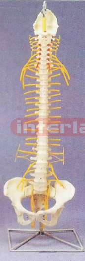
MIDDLE FLEXIBLE VERTEBRAE WITH HANGING LUXURY STAND
MIDDLE FLEXIBLE VERTEBRAE WITH HANGING LUXURY STAND. This detailed, adult spine features nerve branches, vertebral artery, occipital bone, and herniated lumbar disc.Spine can be removed from stand size : 181/2" tall Weight : 5 lbs.

MIDDLE FOOT JOINT FUNCTIONAL MODEL
MIDDLE FOOT JOINT FUNCTIONAL MODEL
