Products
Categories
Educational Equipments
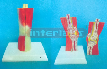
NATURAL SECTION MODEL THROUGH THE KNEE JOINT WITH MUSCLES
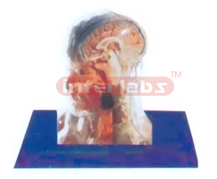
NATURAL TRANSPARENT SKULL ANATOMICAL MODEL WITH ORGANS AND SKELETON
NATURAL TRANSPARENT SKULL ANATOMICAL MODEL WITH ORGANS AND SKELETON
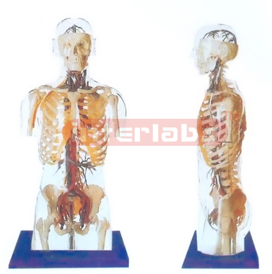
NATURAL TRANSPARENT TORSO MODEL WITH HEAD, HEART, MAIN BLOOD VESSELS AND NERVES IN SKELETON
NATURAL TRANSPARENT TORSO MODEL WITH HEAD, HEART, MAIN BLOOD VESSELS AND NERVES IN SKELETON
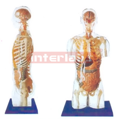
NATURAL TRANSPARENT TORSO MODEL WITH HEAD, ORGANS IN SKELETON
NATURAL TRANSPARENT TORSO MODEL WITH HEAD, ORGANS IN SKELETON
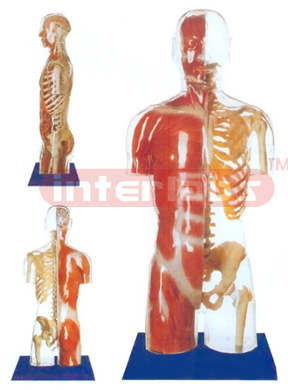
NATURAL TRANSPARENT TORSO MODEL WITH HEAD,SHOWING LEFT SKELETAL SIDE AND RIGHT MUSCLAL SIDE.
NATURAL TRANSPARENT TORSO MODEL WITH HEAD,SHOWING LEFT SKELETAL SIDE AND RIGHT MUSCLAL SIDE.
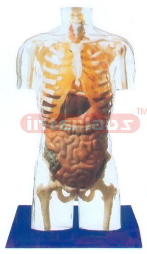
NATURAL TRANSPARENT TORSO MODEL WITH ORGANS & SKELETON WITHOUT HEAD
NATURAL TRANSPARENT TORSO MODEL WITH ORGANS & SKELETON WITHOUT HEAD
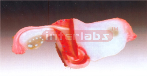
Natural Uterus Model
With the coronal section this model shows:
1. Uterus
2. The cervix is spanided into a supra-vaginal portion and a vaginal portion.
3. The ligaments of the uterus are eight in number.
4. The fallopian tubes
5. The ovaries
Available in Packing: 50 x 35 x 40 cm, 100 pcs/carton, 20 kgs.
1. Uterus
2. The cervix is spanided into a supra-vaginal portion and a vaginal portion.
3. The ligaments of the uterus are eight in number.
4. The fallopian tubes
5. The ovaries
Available in Packing: 50 x 35 x 40 cm, 100 pcs/carton, 20 kgs.
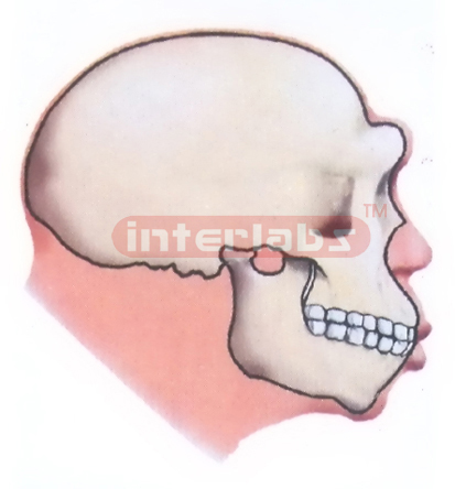
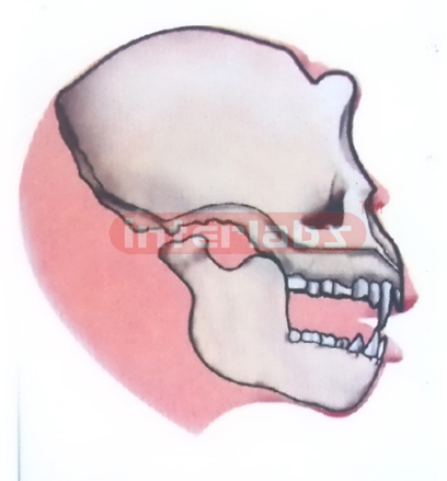
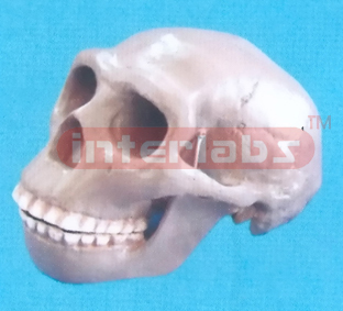
NEANDERTHAL MAN
NEANDERTHAL MAN 150,000 TO 35,000 years ago.
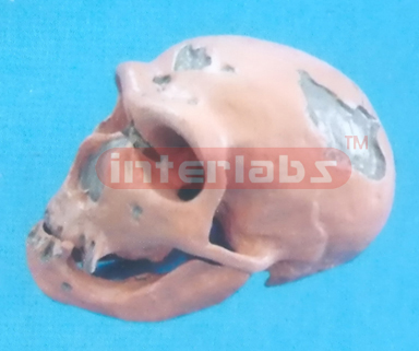
NEANDERTHAL MAN SKULL
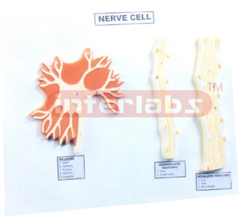
NERVE CELL
Showing different types of Nerve tissue. mounted on a base.
NEURON
Enlarged approximately 2500 times, showing neuron structure clearly based on the latest electron microscopy. Showing medulated nerve fiber. axon, myelin sheath and nodes of ranvier etc., mounted on base with numbered Key Card.
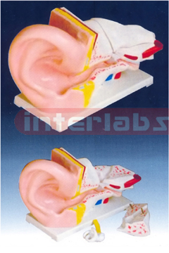
New Style Giant Ear Model
A giant model of the ear, for elementary science classes, shows the three main structural parts of the hearing organ (external ear, middle ear, internal ear) and the position of the equilibrium organ of human body.
1. External ear: Showing the shape of the auricle and the primary features of the external auditory meatus.
2. Middle ear: showing the tympanic membrane, the three auditory ossicles (hammer, anvil, stirrup) and the eustachian tube.
3. Internal ear: showing the vestibule, cochlea and the three semicircular canals of the osseous labyrinth.
This model is dissected into 6 parts. 5 times enlarged. Mouted on plastic base.
Available in Size: 43 x 25 x 15 cm and Packing: 55 x 47 x59 cm, 6 pcs/carton, 18 kgs.
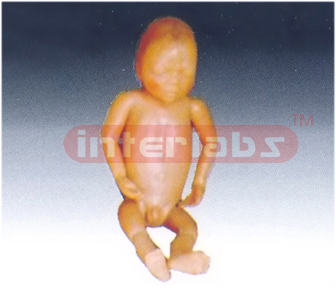
Newborn model (baby modeI) New style newborn model
The size and configuration of this model is almost as same as the normal newborn baby. The head, upper limbs, lower limbs and all joints are flexible. It can demonstrate the characteristic of the newborn baby and all kinds of operations in clinic education: includes different parturition method, newborn baby bath etc. Made of soft pve plastic. Washable. Length: 52cm, head diameter: 34 cm. Available in Packing: 56 x 41 x 73 cm, 10 pcs/carton, 15 kgs.
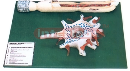
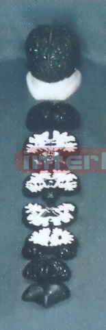
NORMAL LUMBAR COLUMN, ANATOMICAL ANDMAGNIFIED
NORMAL LUMBAR COLUMN, ANATOMICAL ANDMAGNIFIED. The model is designed as an aid for teaching human anatomy and physioliogy course in middle schools and demonstrates the externd features of spinal cord, envelopal by three layers of memb ranes and its relation with spinal nerves within the vertebral canal. 6 times Enlarged Made of
plastic. Size: (including base) 28 x25x49 cm.
Nurse Basic Practice Teaching Model
The basic practice teaching model is designed according to human anatomy and suitable for nursing schools and hospitals for demonstration and practice. It has 5 parts and can demonstrate 15 operations. Part 1: Upper and front part of male body, Part 2: Left arm, Part 3: Male lower abdomen and perineum, Part 4: Hip, Part 5: Female perineum in Packing:
72 x 41 x 43 cm, 1 set/carton, 15 kgs.
72 x 41 x 43 cm, 1 set/carton, 15 kgs.
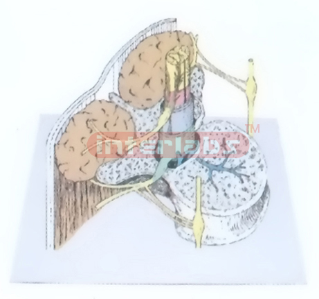
OBIGUE SECTION THROUGH THE FIRST LUMBAR, VERTEBRA SHOING THE SPINAL CORD AND ITS COVERING MEMBRANES
OBIGUE SECTION THROUGH THE FIRST LUMBAR, VERTEBRA SHOING THE SPINAL CORD AND ITS COVERING MEMBRANES

OLDUVAI MAN SKULL
OREOPITHECUS
OREOPITHECUS 5 million to 6 million years ago. (Pliocence Epoch)
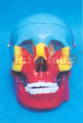
PAINTED ADULT SKULL REPRODUCTION
Cast from a natural bone specimen, this model comtains detailed suture lines, fissares and fossae. Features include a removable calvarium cap, breakaway maxilla and hinged mandible. Each bone is painted a different color for easy identification and the names have been hand lettered. Anatomy mamma and base included. Size : 12.5 X 20 X 18 CM
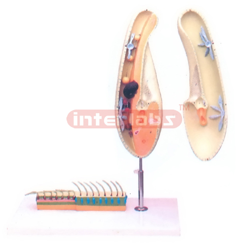
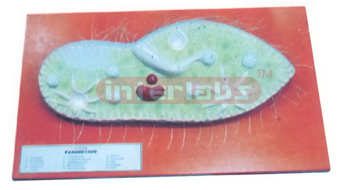
PARAMECIUM
Comprise two detailed, descriptive, accurate and complete models combined in one and mounted on a common base. Provides the overall view of Paramecium, magnified about 1600x. The stand mounting of the model allows convenient viewing by a group of students, even at a distance. The model is sectioned longitudinally, the outer half can be detached to reveal macroand micronuclei, a contractile vacuole, membranelles, myonemes, neuroneme network, food vacuoles, and formation of endo and ectoplasm. The second model, the pellicle enlarged 6,500x, facilitating an upclose view of trichocysts and ciliary bending action illustrating a metachronal wave. The pellicle is mounted directly on the base, while the paramecium is mounted on the stand with numbered Key Card.
PARAMECIUM MODEL
Showing general structure with explanatory notes. On board with numbered key card.
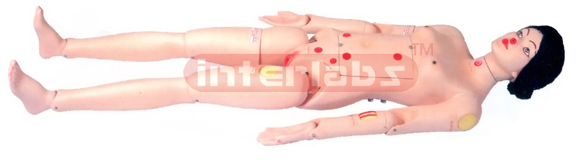
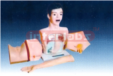
PATIENT CARE SIMULATOR WITH OSTOMY
Provides practice in a wide range of skills from basic nursing through advanced nursing and clinical care: Fully moveable neck, arms and legs, Soft skin of feet, fingers, and toes for added realism, Movable jaw with tongue, Removable dentures, Interchangeable external genital organs allow female and male catherization, Gran, nasal, optic tracheotomy and gastrostomy openings, Transverse colostomy stoma, ileostomy stoma, suprapublic stoma, Detachable at waist ease of storage. Supplied with neck brace.
PEKING MAN SKULL
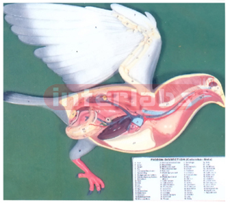
PIGEON DISSECTION
Showing general dissection of a pigeon, an enlarged model mounted on board size 400x480mm approximately. With numbered Key Card.

PIGEON DISSECTION
Showing general dissection of a pigeon in smaller size 320x240mm approximately with numbered Key Card.
PINKISH BRAIN ON LUXURY BASE
PINKISH BRAIN WITH ARTERY ON BASE (8 PCS)
PINKISH BRAIN WITH ARTERY ON BASE (8 PCS). Available in Middle, Small and Little sizes
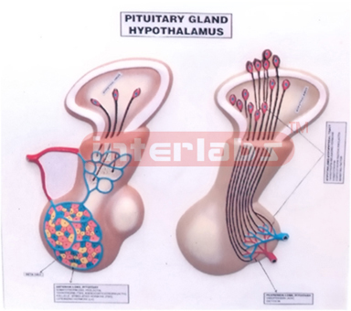
PITUITARY GLAND AND HYPOTHALAMUS
This Model has been designed for the help of Medical Doctors to show the Pituitary Gland and Hypothalamus, mounted on base with numbered Key Card.
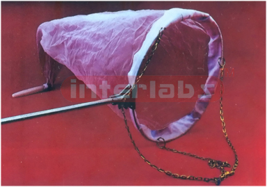
PLANKTON NET
Strong nylon monofilament netting, aperture size 0.1 mm, with tough nylon collar. The mouth of the net is 300mm diameter and the overall length is 900mm. The net supplied complete with frame and handle.
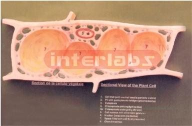
PLANT CELL
Provides clear inside view of the nucleus, chloroplasts, cell wall, and vacuoles of a plant cell, as well as allows exploring the relationship of a cell to its adjacent cells. This simplified model, enlarged 3,000x, is perfect for introducing students to the basic parts of any plant cell. The one-piece model is mounted on a baseboard with numbered Key Card.

PLANT CELL DIVISION MEIOSIS
A newly designed model according to recent concept of chromosome changes from the resting nucleus to the formation of four daughter cells. Complete mounted on board with numbered Key Card.
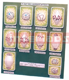
PLANT CELL DIVISION MITOSIS
These models are highly detailed using bright vivid colours, so that they can be clearly seen from a distance by the entire class. A set of 10 models showing all the stages of karyokinesis and cytokinesis from metabolic cells of the formation from one cell to two cells, Mounted on a board with numbered Key Card.
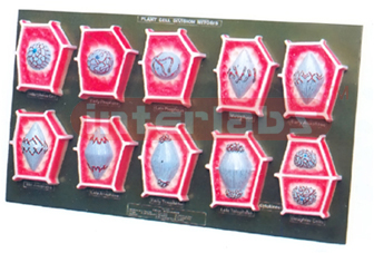
PLANT CELL DIVISION MITOSIS
A set of 10 models showing all the stages of karyokinesis and cytokinesis from metabolic cells of the formation from one cell to two cells shown in simpler and economical version, Mounted on a board with numbered Key Card.
PLANT CELL DIVISION, MEIOSIS
A newly designed model according to recent concept of chromosome changes from the resting nucleus to the formation of four daughter cells. Complete mounted on board with numbered Key Card as a simpler, smaller in size economical model.
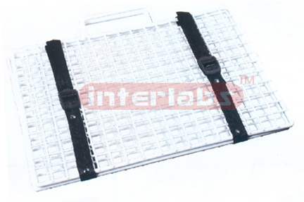
PLANT PRESS
For collecting and pressing plants prior to mounting. The plants are laid between sheet of absorbent paper within the frames and the straps are tightening to apply pressure. The frames are constructed of welded wire rod, incorporating carrying handles. Dip plastic coated. Supplied with straps. Dimensions 425 x 300mm.
PLANT PRESS, WOODEN
Heavy duty brass plants 450 x 300mm, finished with synthetic paint. Two heavy brass chrome plated tightening screws and fly-nuts for uniform pressure. Fly-nuts need not be removed totally to lift the upper plank, but can be tilted on the sides.
PRIMARY HUMAN NERVOUS MODEL
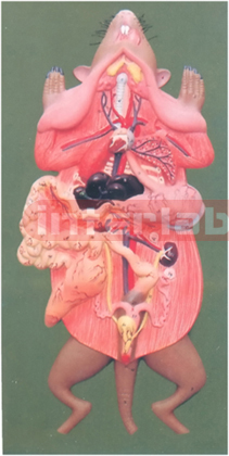
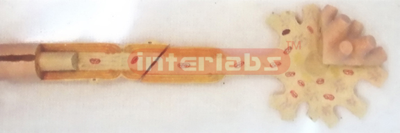
RAT DISSECTION MODEL
This two-part model enlarged 2 1/2 - 3x, is ideal for group study because of its prominent size and enhanced visibility. It shows both atria with auricles, the coronary sulus. the coronary and pulmonary blood vessels, and segments of the esophagus. trachea, aorta, and vena cava. Students can remove the anterior wall to study the internal structures. including the entrance points of the superior and inferior vena cavae and flexible tricuspid, mitral. and aortic valves. The model is attached on a stand, which represents a portion of the pericardium resting in its natural position on the diaphragm. The accompanying key identifies 59 numbered features on the model. Size S"x 7" x 10" [LxWxH]

RHOOESIAN MAN SKULL
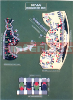
RIBONUCLEIC ACID (RNA)
Model showing RNA molecule, DNA template molecule of chromosome, role of messenger and transfer RNA colour key. Mounted on board with numbered Key Card.
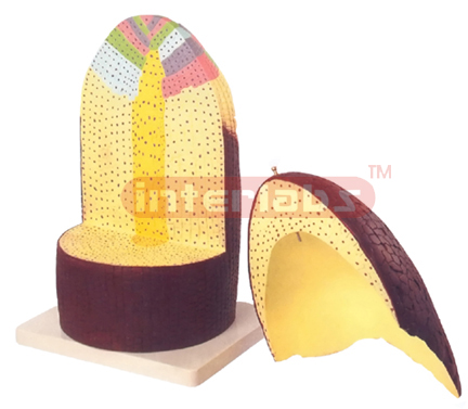
ROOT
Enlarged about 300 times, durable plastic, showing in longitudinal and transverse section, crown of removable. Dissectible in two part. Mounted on base with numbered Key Card.
ROOT TIP ANATOMY
Showing LS and TS of a root tip with root cap maturation region and root hairs etc. A threedimensional model, mounted on stand with numbered Key Card.
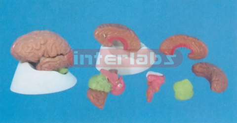
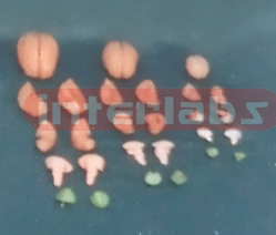
SAGITAL PLANE BRAIN MODEL (7 PCS )
SAGITAL PLANE BRAIN MODEL (7 PCS )
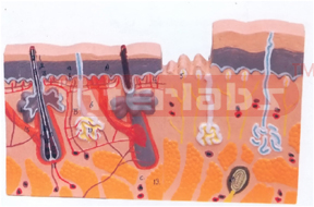
SECTION OF HUMAN SKIN
A full size model, showing section through 3 layers of the skin, showing hair follicles with sebaceous, sweat glands, receptors, nerves and vessels, 70 times enlarged, but mounted in upright position, a very detailed model with numbered Key Card.
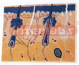
SECTION OF HUMAN SKIN
This relief model shows a section through the three layers of the haircovered skin of the head. Representation of hair follicles with sebaceous glands, sweat glands, receptors, nerves and vessels. Delivered on base with numbered Key Card.
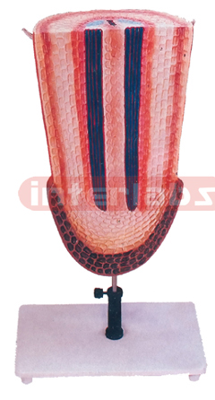
SECTION THROUGH THE STEM OF A YEAR OLO DICOTYLE PLANT
Lime tree, Tilia sp., somewhat simplified, single piece model, enlarged approx. 125 times. Mounted on a base with explanatory key card.
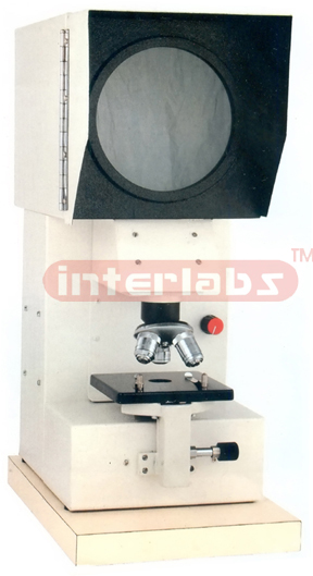
SENIOR PROJECTION MICROSCOPE
The Senior Projection Microscope is used for Textile Industry, fibre structure and thickness of woolen fibers. This Latest and most advanced projection microscope with Binocular head with switch over system facilitating observation of specimen through binocular head as well as on the screen.
Features
· Comes with a 200mm graduated Observation Screen, built-in Fresnel lens, loop and arrow arrangement and Co-axial mechanical stage of size 135 x 120mm.
· Provides magnification from 125x to 1000x.
· Illumination provided by a Halogen Lamp 12V, 100W, workable on 220V / 110VAC.
· Quadruple revolving nosepiece.
Optical Combinations
Eyepieces WF 10 x paired
Objectives 5x, 10x, 20x and 40x
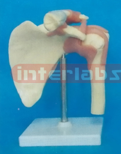
SHOULDER JOINT-FUNCTIONAL MODEL